Carotid Artery Disease shows no symptoms until a transient ischemic attack or stroke happen.
Depending on your medical history, your doctor will perform a thorough physical examination and order an imaging test of the carotid arteries to rule in (or rule out) Carotid Artery Disease.
In case of a confirmed transient ischemic attack or stroke imaging of the carotid arteries will be done immediately.
Your doctor may use a stethoscope to listen to the arteries in your neck.
If an abnormal sound, called a bruit, is heard it may reflect turbulent blood flow, indicative of a narrowed carotid artery.
However, bruits are not always present when there are blockages, or they may be heard even when the blockage is minor.
Be sure to let your doctor know if you have had any symptoms, such as those listed above. Imaging tests include:
Carotid Ultrasound
Carotid Ultrasound: It is simple, fast, non-invasive and accurate in the diagnosis of Carotid Artery Disease.

It can be used for screening or surveillance of known Carotid Artery Disease.
Computed Tomography Angiography (CTA)
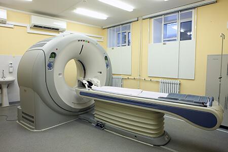
Computed Tomography Angiography (CTA): It is a very accurate study for the diagnosis of Carotid Artery Disease.
It can give high resolution 3D pictures of the carotid arteries and more information for their intracranial portions, as well as for the brain.

This comes at the cost of the need for radiation and intravenous dye (iodinated contrast) that may be harmful for the kidneys, especially in patients with impaired renal function.
CT scans are not ordered routinely, unless there is a confirmed ischemic event or your vascular surgeon plans surgery (carotid endarterectomy or stenting).
Magnetic Resonance Angiography (MRA)
Magnetic Resonance Angiography (MRA): It can be as accurate as the CTA, doesn't require radiation and if intravenous contrast is used (gadolinium) it is less harmful for the kidneys.

An MRA can often detect even small strokes in the brain.
Drawbacks are that it may be time consuming, it is expensive and it may be contraindicated in patients with claustrophobia or metallic implants.
Traditional Angiography
Traditional Angiography: It is an invasive imaging study done in a surgical environment as it requires arterial access through a groin puncture.
It requires radiation and intravenous contrast and it may be used to assist your vascular surgeon's operative plan.

In contemporary practice, it is not routinely recommended for diagnosis.
It will eventually be used as part of your treatment if carotid stenting has been decided.


